3D bioprinting is the process of building a three-dimensional structure layer by layer out of living cells and biological materials, usually cells suspended in a printable gel called bioink, to make tissue models, implants and eventually replacement organs. If you have followed the headlines, it sounds like a science fiction printer that spits out a kidney on demand. That is not where the technology is.
This guide covers bioprinting basics explained simply, from the cells and the gel to the printer head, the uses researchers put these structures to, and the honest reasons whole printed organs are still out of reach. No biology background assumed.
The short version: bioprinting is ordinary additive manufacturing with a living twist. A printer places cell-laden gel one layer at a time, the gel is locked into a solid scaffold, and the cells are then fed in a bioreactor so they can grow, connect and mature.
Table of Contents
- What Is Bioprinting?
- How Does Bioprinting Work?
- Bioprinting Basics Explained Simply: From Cells to Scaffolds
- What Materials Are Used in Bioprinting?
- Which Bioprinting Technologies Are Used?
- Why Do Bioprinted Structures Need Scaffolds?
- What Is Bioprinting Used For Today?
- Bioprinting Basics: Key Benefits and Limitations
- How Is a Bioprinted Sample Evaluated?
- Can You Learn Bioprinting as a Beginner?
- Frequently Asked Questions
- Is bioprinting the same as 3D printing a human organ?
- What is the difference between 3D bioprinting and 4D bioprinting?
- Do all bioprinters use living human cells?
- Why are fully functional printed organs still difficult to make?
- Is bioprinting used in routine medical treatment today?
- What is the most accessible way for beginners to learn bioprinting?
- Conclusion: Where to Start with Bioprinting
What Is Bioprinting?
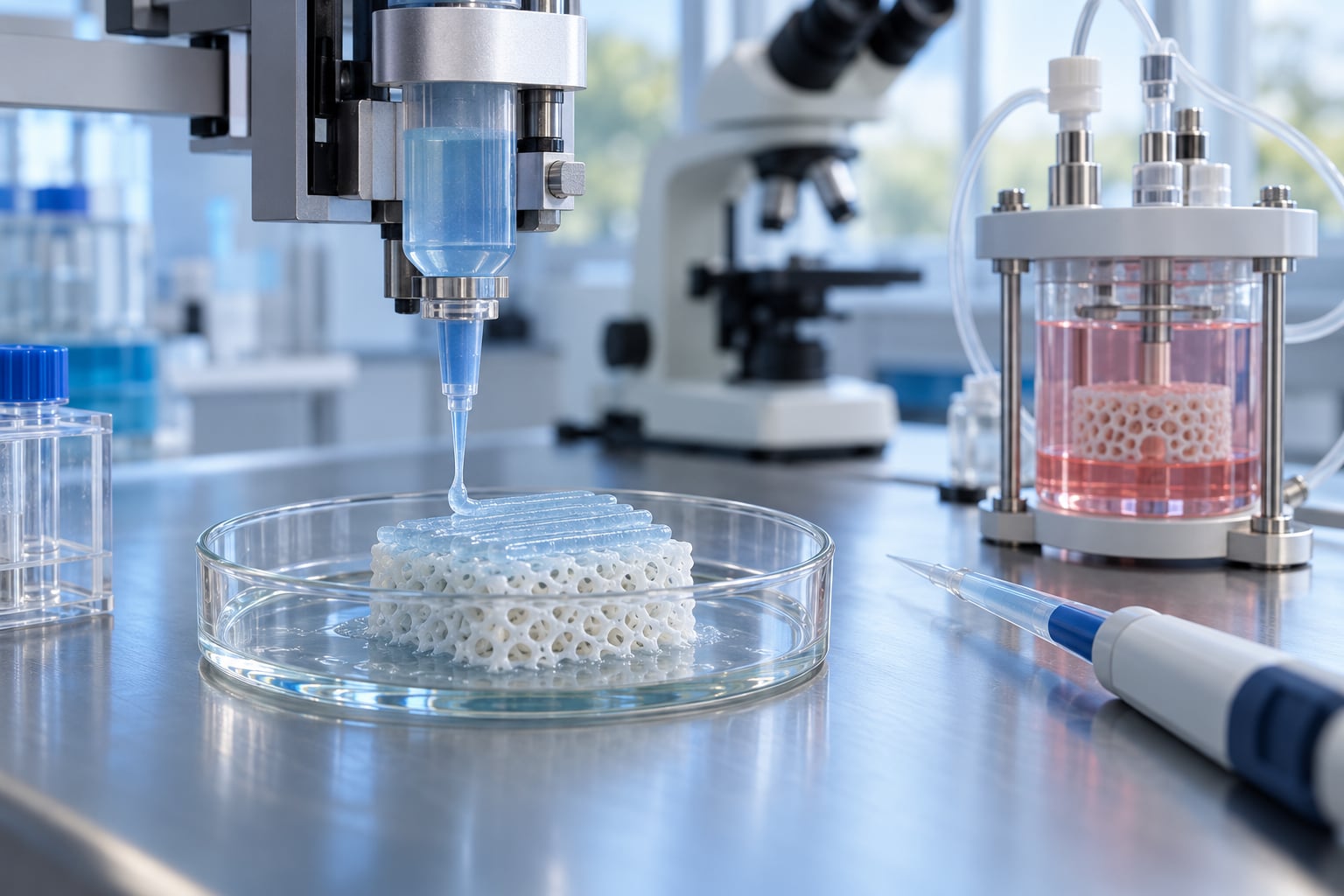
Bioprinting is a form of additive manufacturing that builds a physical object by adding material on top of material, layer by layer, until a three-dimensional shape exists. The unusual part is what gets added. Instead of melted plastic or powdered metal, a bioprinter deposits cells, molecules taken from living tissue, or both, suspended in a printable gel.
Every bioprinting job has three ingredients. You need cells, or a cell-free stand-in for them. You need bioink, the printable material that carries those cells and holds its shape once deposited. And you need a printer, which is often a modified extrusion machine sitting inside a temperature-controlled enclosure.
That last part is where the difference from your desktop printer becomes obvious. A standard machine melts filament at around 200 degrees Celsius. Cells cannot survive that. A bioprinter works near body temperature, keeps everything sterile, and often protects the material from being knocked about by the moving parts.
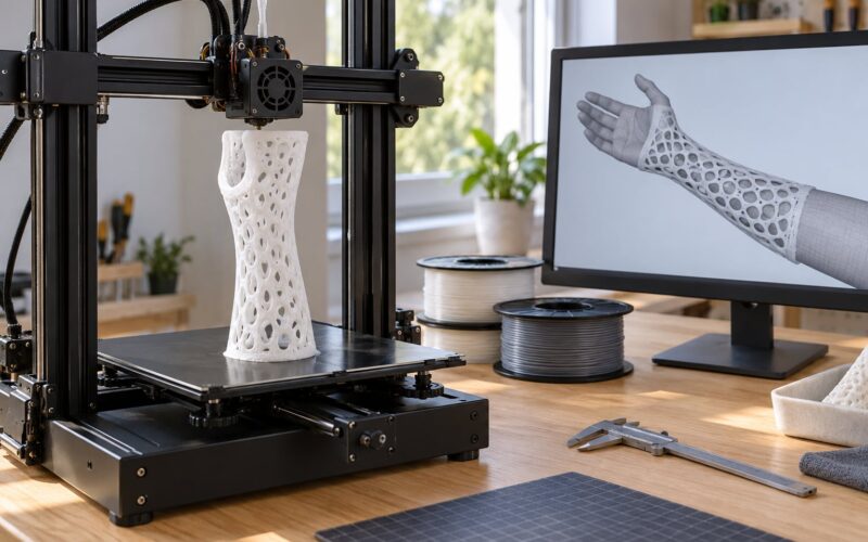
The distinction also depends on what comes out. Printing a plastic model of a jaw from a CT scan is medical 3D printing, and it is genuinely useful in surgery, but it is not bioprinting. Nothing in it is alive. Bioprinting is specifically about building something that contains living material or a biological scaffold meant to host it.
| Feature | Ordinary 3D printing | 3D bioprinting |
|---|---|---|
| Material | Plastic, resin, metal, ceramic | Cells, hydrogels, decellularized tissue, biological composites |
| Working temperature | Often 200 degrees Celsius or more for thermoplastics | Near body temperature, usually 20 to 37 degrees Celsius |
| Sterility | Not required | Full sterile workflow throughout |
| Goal | A finished solid part | A structure that stays alive and keeps changing |
| After printing | Remove supports and use it | Crosslink, then mature in a bioreactor for days to weeks |
| Main constraint | Melting point and printer frame | Cell viability and the need for nutrients and blood supply |
How Does Bioprinting Work?
The workflow is closer to cooking than to manufacturing. Every stage depends on the one before it, and skipping a step ruins the result. Here is the standard sequence, from a medical scan to a living tissue model.
- Design the shape. Researchers start with a digital model. For patient-specific work this comes from a CT or MRI scan, which is converted into a 3D file, then into a toolpath the printer can follow.
- Grow and prepare the cells. Cells are expanded in culture so there are enough of them to print with. Some come from a donor biopsy; others are induced pluripotent stem cells, which are adult cells rewound to an earlier, more flexible state and then grown into the tissue type needed.
- Mix the bioink. Cells are suspended in the gel at a chosen density. Printability and cell comfort pull in opposite directions, and finding the balance is most of the practical skill involved.
- Print the construct. The printer deposits the bioink in a controlled pattern, one layer at a time, while keeping the environment cool, humid and free of contamination.
- Crosslink the structure. A trigger such as ultraviolet light, calcium ions, temperature or an enzyme locks the gel into a solid scaffold so it holds its printed shape.
- Mature it in a bioreactor. The construct goes into a vessel with nutrients, oxygen and sometimes mechanical stimulation, and is left to grow for days or weeks before testing.
Bioprinting Basics Explained Simply: From Cells to Scaffolds
A useful picture is baking a jelly cake with raisins in it. The jelly is the bioink, the raisins are the cells, and the heat that sets the jelly is crosslinking. Once it comes out of the tin, the cake still has to rest in a cool place for the fruit to soften and release colour. That resting stage is the bioreactor.
Each piece has its own job. Cells do the biological work: they multiply, lay down their own support material, and eventually become the tissue you were aiming for. The biomaterial does the structural work, giving the cells something to hold on to in three dimensions. The scaffold is the framework itself, and it may fade away on a schedule or stay for good. The printing parameters, meaning speed, pressure, temperature, layer height and nozzle size, decide whether the whole thing holds together.
That analogy also explains the hardest trade-off. Make the gel stiff enough to hold a detailed shape and the cells feel trapped inside something rigid. Make it soft enough to live in and it slumps the moment the nozzle moves away. Most of the real research goes into that squeeze.
What Materials Are Used in Bioprinting?
Bioprinting basics explained simply come down to trade-offs rather than one perfect material. Researchers mix groups to get what a specific tissue needs, and the same base gel can behave very differently once it is blended.
| Material group | Cell compatibility | Printability | Stability | Typical use |
|---|---|---|---|---|
| Living cells (primary, stem cell lines, iPSCs) | Self, by definition | Must be kept in suspension | Needs constant nutrients | Functioning tissue models and implants under study |
| Natural hydrogels (collagen, fibrin, alginate, hyaluronic acid) | High, they resemble body tissue | Often too soft to print alone | Varies, some need constant support | Soft tissue, skin, cartilage and wound models |
| Synthetic polymers (GelMA, PEGDA, Pluronic F-127) | Mixed, tunable by chemistry | Good, adjustable viscosity | Often predictable and repeatable | Structured models and drug-testing platforms |
| Decellularized tissue (dECM) | High, it is native matrix | Difficult, dense and fibrous | Degrades on a biological schedule | Soft tissue scaffolds that encourage natural growth |
| Composite bioinks | Tuned per formulation | Best of several properties | Balanced | Realistic multi-tissue models where one gel will not do |
Hydrogel deserves a note, because it comes up constantly. A hydrogel is a network of long molecules that holds water. Skin, cartilage and much of the eye are naturally hydrogels, which is why water-based gels work so well as a starting scaffold.
Decellularized tissue is the other idea worth understanding. Take an organ, wash out every living cell, and what remains is the extracellular matrix, the fibrous web the cells were sitting in. That web already has the right architecture for the tissue, so printing it can give cells a much better starting template than anything built from scratch.
Which Bioprinting Technologies Are Used?
Five approaches cover most of the work being published. They differ mainly in how the material gets from the cartridge to the build platform.
| Method | Deposition | Useful outputs | Main advantages | Key limitations |
|---|---|---|---|---|
| Extrusion printing | Pneumatic, piston or screw pressure pushes gel through a nozzle | Bulk tissue, scaffolds, sacrificial channel networks | Handles viscous inks and real cells, works at large scale | Resolution in the hundreds of micrometres, cells feel shear stress in the nozzle |
| Inkjet or droplet printing | Drops ejected on demand, like an office printer | Fine patterns and multiple cell types side by side | High precision, gentle on cells, many nozzles at once | Needs thin inks with low viscosity, clogs easily |
| Direct bioprinting and in situ printing | Deposition straight onto the patient or the wound site | Skin grafts, cartilage repair during surgery | No handling, no freezing, matched to tissue contours | Sterility and time pressure in an operating theatre |
| Laser-assisted printing | Laser energy pulls material from a donor layer onto the substrate | High-resolution patterns from several cell types | Very fine resolution and high cell density | Specialised equipment and slow build speeds |
| Light-based and volumetric printing | Projected light solidifies a whole 3D volume at once | Complex 3D shapes in seconds, organ-on-a-chip devices | Fastest route to intricate forms, smooth surfaces | Limited to transparent resins and cells exposed to light stress |
One technique is worth naming because it keeps showing up in papers. FRESH, short for freeform reversible embedding of suspended hydrogels, prints hydrogel inside a temporary support gel that later melts away. It exists because a soft material cannot hold its own shape in mid-air, and this trick lets researchers print structures that would otherwise collapse.
Why Do Bioprinted Structures Need Scaffolds?
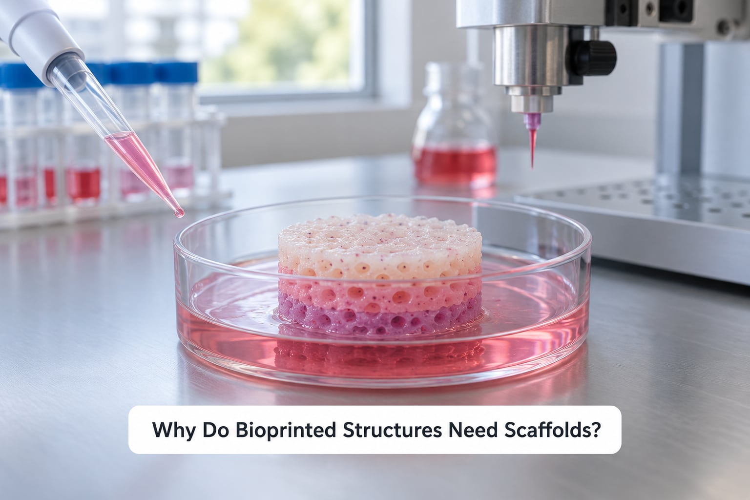
Cells behave badly on their own. Suspended in a dish they form flat clumps, and flat clumps do not grow into anything resembling a bone or a heart wall. Human tissues are three-dimensional, and cells need physical guidance to organise themselves into that shape.
A scaffold is that guidance. It gives cells a three-dimensional framework to attach to, hold them near each other so they can signal, and set the spacing between them so growth happens in the right direction. Without it you get a pile of cells. With it you get a structure.
Any explanation of bioprinting basics explained simply keeps returning to this one problem. There are two categories of scaffold, and the difference between them matters more than it sounds. A cell-free scaffold, often called acellular, is printed on its own with no living cells present. It is a ready-made structure for later seeding, or simply a model for testing. A cell-bearing construct has living cells deposited inside the scaffold during printing, so the cells and the framework are built together.
Scaffolds also come with different end dates. Some are sacrificial, meaning the cells digest or replace the printed structure as they grow, which is how most soft tissue ends up living in its own matrix. Others are designed to persist, such as a rigid printed frame for a bone-shaped implant. The right choice depends entirely on whether you want the scaffold to disappear or stay.
What Is Bioprinting Used For Today?
Most bioprinting today is research work, and the most established commercial use has nothing to do with implants. It has to do with testing drugs and chemicals on human cells that were grown in the lab rather than on animals. Companies in cosmetics, pharmaceuticals and toxicology use printed tissue models because regulators in Europe and the United States increasingly expect product safety data built on human-relevant methods.
What bioprinting is used for in research and medicine today falls into a few clear groups.
- Drug and toxicity testing. Printed liver, kidney and cardiac tissue models give researchers a human response to test against, which catches problems earlier than animal work alone.
- Disease and tumour modelling. Researchers print tissue that mimics a tumour environment to study how cancer cells invade and how drugs slow them down.
- Organ-on-a-chip devices. Microfluidic chips holding small printed tissues model the interaction between organs, such as a gut barrier with immune cells moving through it.
- Surgical planning and rehearsal. Patient-specific printed models let a surgeon practise a difficult operation before the patient is on the table.
- Teaching. Printed anatomy models are appearing in medical schools, including tissue-like structures students can dissect or study repeatedly.
- Early implant work. The closest clinical results to date are narrow and structural: printed tracheal splints for infants with airway collapse, printed skin for burns and severe wounds, cartilage repair patches, and corneal tissue for vision restoration trials.
- Regenerative medicine research. Scaffolds seeded with a patient’s own cells are being investigated for bone and soft tissue repair, mostly in laboratory and animal studies so far.
The honest summary is that bioprinting as a drug-testing and tissue-modelling tool is real and commercial. Bioprinting as a source of transplant organs is still research. Anything telling you otherwise is jumping ahead by decades.
Bioprinting Basics: Key Benefits and Limitations
Every benefit below comes with a catch, and the catch is the interesting part.
| Benefit | What it means | Limitation that comes with it |
|---|---|---|
| Control over structure | You decide where every cell sits instead of hoping a moulded shape works | Resolution is far coarser than plastic printing, usually hundreds of micrometres |
| Patient-specific models | A scan of one person becomes a model of that same person | Each model takes time, so it suits planning rather than mass production |
| Fewer animal tests | Human cells can replace some early safety screening | Human models miss reactions that only a whole living animal shows |
| Reproducibility | The same digital file makes the same construct every time | Living cells drift from batch to batch, so “same” is never exact |
| Fewer donor transplants | Grafts grown from a patient’s own cells dodge immune rejection | Regrowth takes weeks, which suits planned surgery rather than emergencies |
| Lower material waste | Additive manufacturing only uses what it prints | Bioink is expensive and sterile, so waste is not the limiting factor |
Three problems run underneath all of it. The first is vascularisation. Cells need to sit within roughly 100 to 200 micrometres of a blood vessel to get enough oxygen and nutrients. Beyond that they die. A cell clump a few millimetres across is already outside that range, and no printer yet builds the branching capillary network a real organ needs.
The second is maturation. A printed construct is not a finished tissue. It has to live in a bioreactor for days or weeks, sometimes with mechanical stimulation, to develop the strength and organisation of the tissue it is meant to represent.
The third is oversight. A printed implant that carries living cells sits somewhere between a device, a biological product and a tissue graft, and it has to be judged against all of those rules at once.
How Is a Bioprinted Sample Evaluated?
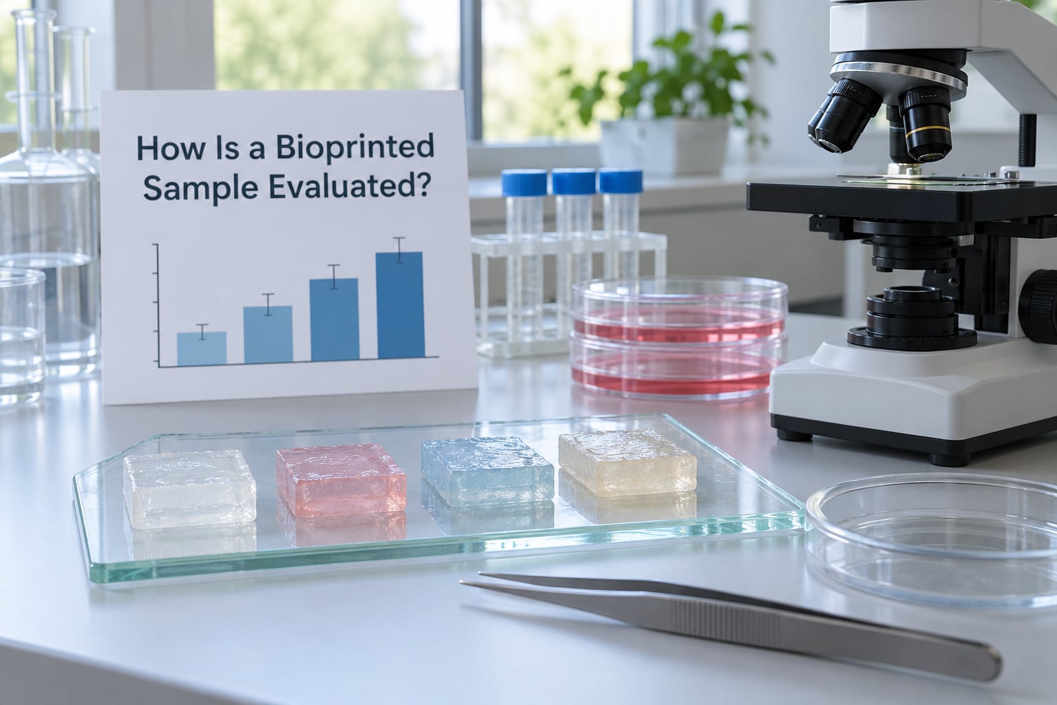
Print something and it is only the start. A construct has to be measured, and the same checklist gets used whether it is a first experiment or a clinical programme.
Cell viability comes first, measured with live-dead staining before and after printing. Above 85 percent is a common target, because there is no point building a beautiful structure out of dying cells.
Printability is checked by asking whether the shape survived the process at all. Pores collapse, layers smear or the sample tilts, and the run is usually discarded.
Layer adhesion is tested by pulling or peeling. If the layers separate under light force, the whole construct fails no matter what the images show.
Mechanical behaviour is measured by compression or tensile testing. A cartilage construct should feel springy and a bone construct should resist load, so matching the target tissue is the actual criterion.
Porosity and degradation are tracked over time. Pores let nutrients and waste move through a structure, and the degradation rate has to match how fast new tissue replaces the scaffold.
Differentiation answers the biggest question: are the cells still just cells, or have they started behaving like the tissue they were meant to become?
Sterility gets verified with no short cuts, because a single contaminant ruins months of work.
Can You Learn Bioprinting as a Beginner?
Yes, and the entry points are more open than they were five years ago. What you cannot do safely or cheaply is anything involving live human cells, and that boundary is worth respecting.
Bioprinting basics explained simply for beginners start with cell-free work. Printing a hydrogel scaffold in alginate, gelatin or a Pluronic-based gel teaches you slicing, extrusion pressure, layer height and how bioinks behave without any biosafety requirements. Models of bone, cartilage, nerve pathways and blood vessels all work this way, and they appear in student projects and hobby forums constantly.
Beyond that, tissue engineering courses at universities and community colleges are the standard next step, usually covering cell culture and standard lab technique. Open-source microscope and image-processing tools let you document a sample properly without expensive imaging gear, and open hardware designs for extrusion bioprinters have made the machines themselves buildable at a fraction of commercial prices.
Working with living cells needs something else entirely: a clean room, an incubator, culture hoods and trained supervision. Those facilities are not something to improvise, and most people who advance into live-cell research do it inside a university or hospital lab rather than at home.
Frequently Asked Questions
Is bioprinting the same as 3D printing a human organ?
No. Bioprinting means building a structure out of biological material, such as cells suspended in a printable gel, and most real applications are tissue models for research rather than transplantable organs. Researchers can print small organ-like models for drug testing, but a working kidney or liver with its own blood supply does not exist yet.
What is the difference between 3D bioprinting and 4D bioprinting?
3D bioprinting builds a structure that is fixed once it is printed. 4D bioprinting adds change over time, usually by using a material that responds to heat, moisture, light or pH, so the printed shape moves, bends or unfolds after printing. The technique is promising for self-shaping scaffolds and soft actuators, but it remains early research.
Do all bioprinters use living human cells?
No. Some print cell-free scaffolds and biomaterial structures for testing or for later seeding with cells. Others print human cells, cells from animals, or entirely biological materials such as decellularized tissue matrix with the original cells removed. Printed models that are meant to stay alive need cells and a bioreactor to feed them afterwards.
Why are fully functional printed organs still difficult to make?
The main obstacle is blood supply. Cells need oxygen and nutrients within roughly 100 to 200 micrometres of a blood vessel, and beyond that distance they die. No printer yet produces the dense branching capillary network every organ depends on. Layer resolution, long maturation times and regulatory review add further delays.
Is bioprinting used in routine medical treatment today?
It is used routinely in research and drug testing, where printed human tissue models are standard practice. A small number of clinical applications have reached patients, including printed tracheal splints for infants, skin grafts for severe wounds and corneal tissue in clinical trials. Whole printed organs are not available as treatment.
What is the most accessible way for beginners to learn bioprinting?
Start with cell-free hydrogel scaffolds printed on an entry-level extrusion bioprinter. You can learn slicing, extrusion pressure, layer height and bioink behaviour without cell culture facilities or biosafety requirements. From there, tissue engineering courses and university lab projects are the usual route into anything involving live cells.
Conclusion: Where to Start with Bioprinting
Bioprinting is additive manufacturing with cells in the mix. Cells are suspended in a printable gel, deposited layer by layer, locked into a scaffold, and matured in a bioreactor so they can grow into tissue that behaves like the real thing.
The realistic picture matters here. Drug testing and disease modelling are commercial uses running today, and a small set of structural implants have reached patients. Whole printed organs remain research, held back mostly by the fact that cells cannot survive more than a fraction of a millimetre from a blood vessel.
One clear first step: learn the vocabulary first. Cells, bioink, scaffold, crosslinking, bioreactor and cell viability explain almost every headline you will meet. That is bioprinting basics explained simply, and once those words are familiar, read any bioprinting claim with the vascularisation limit in mind, because that single fact separates most real advances from the ones that are years away.